
Researchers at Pitt have produced the most detailed image to date of a bacteriophage–phage for short–that has allowed them to see for the first time the structural makeup of the part of the phage that directly attaches to its target Mycobacterium cell.
There are about 1031 phages on Earth–about one trillion for every grain of sand–and they have been evolving for billions of years. Despite all the variation this long history has given rise to, they almost all share similar components: a capsid, a tail tube, and a tail tip.

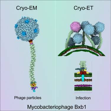
Publication: Structure and infection dynamics of mycobacteriophage Bxb1
“Mycobacteriophage Bxb1 is a well-characterized virus of Mycobacterium smegmatis with double-stranded DNA and a long, flexible tail. Mycobacteriophages show considerable potential as therapies for Mycobacterium infections, but little is known about the structural details of these phages or how they bind to and traverse the complex Mycobacterium cell wall. Here, we report the complete structure and atomic model of phage Bxb1, including the arrangement of immunodominant domains of both the capsid and tail tube subunits, as well as the assembly of the protein subunits in the tail-tip complex. The structure contains protein assemblies with 3-, 5-, 6-, and 12-fold symmetries, which interact to satisfy several symmetry mismatches. Cryoelectron tomography of phage particles bound to M. smegmatis reveals the structural transitions that occur for free phage particles to bind to the cell surface and navigate through the cell wall to enable DNA transfer into the cytoplasm.”
